
Fig. 4.
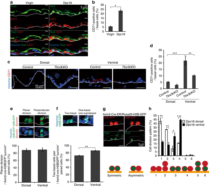
Axin2+ IFE stem cells generate Tbx3+ basal cells through planar-oriented symmetric/asymmetric cell division. a Immunofluoresence of Tbx3 (green), CD71 (red) and Hoechst (blue) on ventral skin from virgin and 16 dpc mice. b Quantification of CD71+ cells in IFE basal layers. c Immunofluoresence of CD71 (red) and Hoechst (blue) on tamoxifen-treated dorsal and ventral skin from 16 dpc control and Tbx3 cKO mice interbred with genetically unaltered males. d Quantification of CD71+ cells in IFE basal layers. e, f Immunofluoresence of GFP (green), survivin (red) and Hoechst (blue) on ventral skin from 16 dpc Axin2-Cre-ER/Rosa26-H2B-GFP mice. Quantification of planar-oriented IFE basal cell division in Axin2-Cre-ER labelled-survivin+ cells (e) and the two basal cell pairs in Axin2-Cre-ER labelled-survivin− cells (f) are shown on the bottom. g Typical images of planar-oriented symmetric and asymmetric division of Axin2-Cre-ER-labelled IFE basal cells. h Quantification of the division pattern of Axin2-Cre-ER-labelled IFE basal cells in dorsal and ventral skin at 16 dpc. b, d–f, h Data represent the mean ± s.e.m. of averages of three independent experiments (n = 3). > 500 cells (b), > 200 cells (d), > 40 cells (e), > 150 cells (f), > 50 cells (h) were analysed to calculate the average in each experiment; *P < 0.05, **P < 0.01, ***P < 0.001, analysed by the Dunnett’s multiple comparison test (d), two-tailed t-test (b, e, f), or Tukey’s multiple comparison test (h). a, c, e, f, g White-dotted lines represent basement membranes. Experiments were repeated three times with three mice for representative images