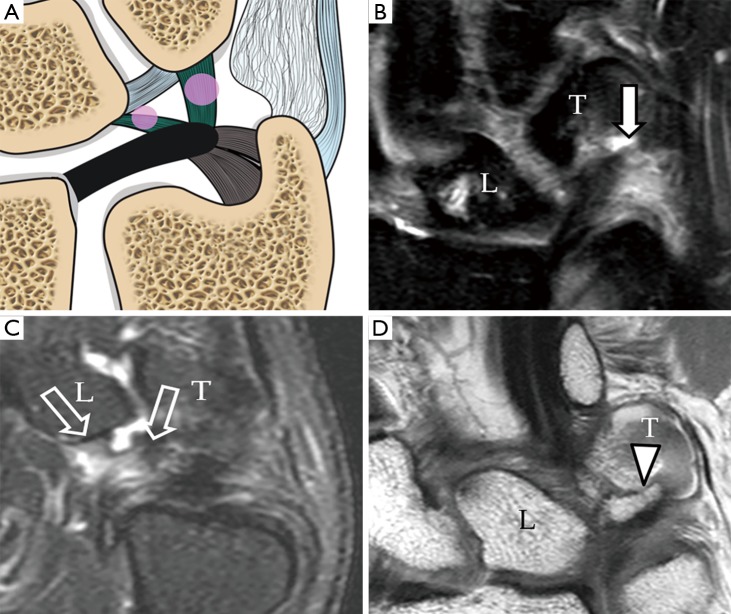
Figure 11.
Type 1C tear. (A) Schematic drawing showing tear of the ulnolunate and ulnotriquetral ligaments (pink circles); (B) proton density fat suppressed coronal MRI image shows severe oedematous change with thickening at the ulnotriquetral ligament (short solid arrow) consistent with a intrasubstance partial tear of this ligament; (C) proton-density fat suppression coronal MRI image of another patient showed severe oedematous change with thickening at the ulnolunate ligament (short block arrows) attaching to proximal lunate (L) consistent with an intrasubstance partial tear; (D) proton-density coronal MRI image of another patient shows avulsion fracture of proximal triquetrum (T) at the attachment of the ulnotriquetral ligament (solid arrowhead).

