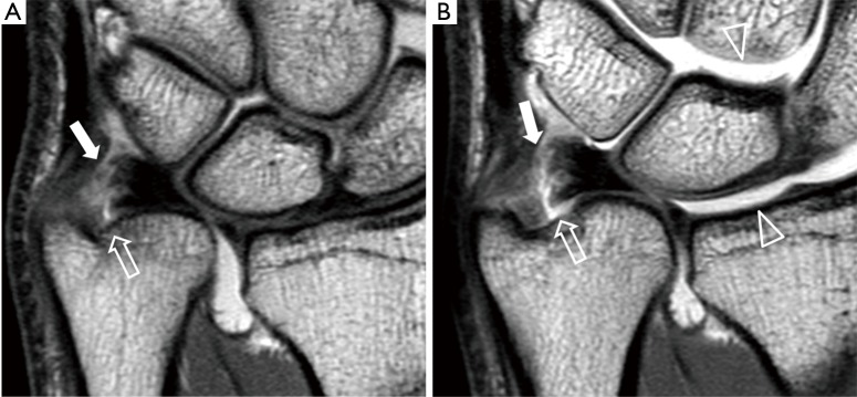
Figure 20.
Comparison of TFCC visualization using non-traction MRI (A) and traction MRI (B). Before traction, there is a tear depicted at the foveal attachment (block arrow) of the TFCC but the styloid attachment appears intact (solid arrow). After traction, discontinuity at the styloid attachment can be fully appreciated (solid arrow). The radiocarpal and the mid-carpal joints are widened following traction (arrowheads). TFCC, triangular fibrocartilage complex.

