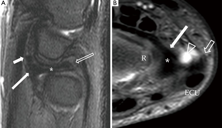
Figure 6.
Normal appearance of the TFCC on sagittal T2W fat-suppressed (A) and axial proton-density fat suppressed (B) images. (A) The TFC (asterisk) is concave in appearance on a sagittal view. On the volar side, the TFC is attaching to the volar radioulnar ligament (vRUL) (long solid arrow) and more distally through the ulnotriquetral ligament (short solid arrows). On the dorsal side, the TFC is attaching to the dorsal radioulnar ligament (block block arrow). These structures are not seen as distinct structures on MRI when they are normal; (B) the TFC (asterisk) is barely seen on axial image. On the volar side, the TFC is attaching to a low signal linear structure representing the volar radioulnar ligament (vRUL). On the radial side, it attaches to the distal radial cartilaginous rim (R). On the ulnar side, the attachments are very difficult to be appreciated at this level. However, the location of the distal ulnar styloid process (short block arrow) and the ECU can be appreciated on this image. Fluid within the pre-styloid recess is seen (arrowhead). TFCC, triangular fibrocartilage complex; ECU, extensor carpi ulnaris.

