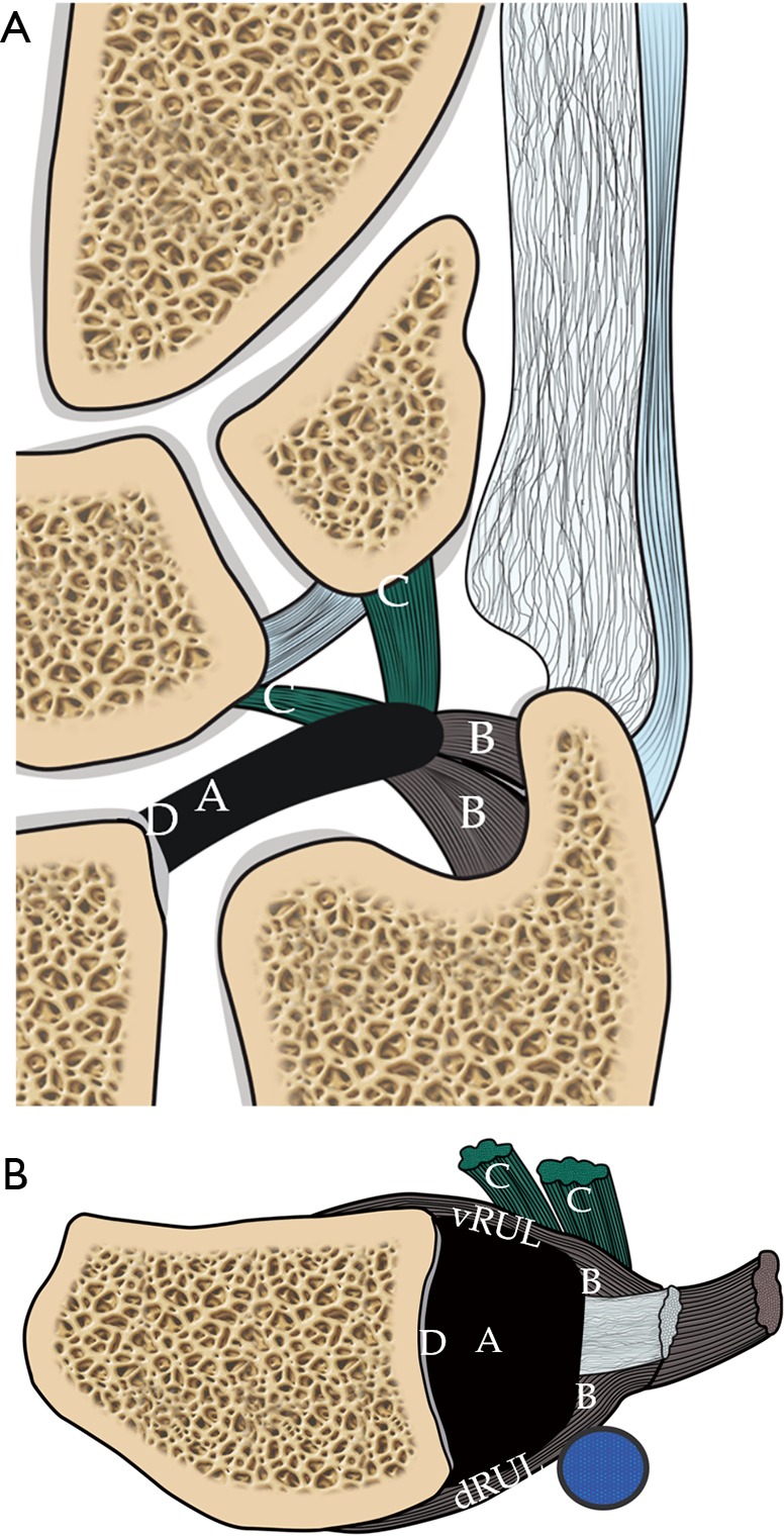
Figure 7.

Schematic diagram showing the Palmar classification of TFCC tears. (A) Coronal view shows the different types of tear correspond to the location of the tear. Type 1A is central TFC perforation. 1B, peripheral ulnar side TFCC tear (± ulna styloid fracture). 1C, distal TFCC disruption (disruption of distal UC ligaments) and 1D, radial TFCC disruption (± sigmoid notch fracture); (B) axial view shows the different tear locations. A tear on the volar and dorsal sides of the TFC involving the volar (vRUL) or dorsal radioulnar ligament (dRUL) is not included in the Palmar classification. TFCC, triangular fibrocartilage complex.
