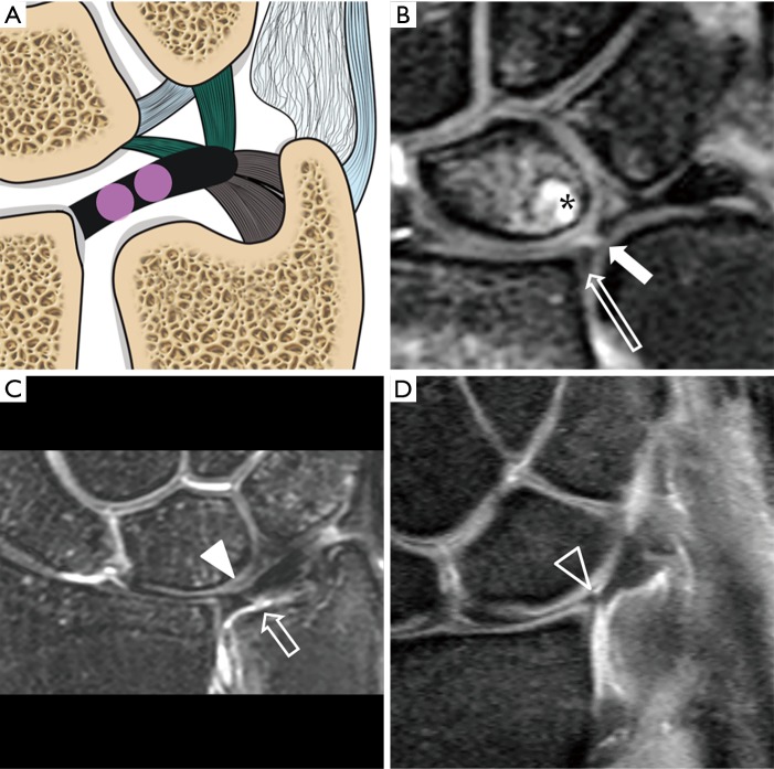
Figure 8.
Type 1A tear. (A) Schematic drawing showing the tear (pink circle) at the central or paracentral part of the TFCC. Proton density fat suppression Coronal MRI images showing (B) full thickness tear with a small gap filled with fluid (short solid arrow). There is a small remnant of TFC at the radial attachment (long block arrow). A small subchondral cyst is at the proximal ulnar side of the lunate bone (asterisk); (C) partial thickness tear at the undersurface of the TFC (short block arrow). The distal surface of TFC is intact with the contour preserved (solid arrowhead); (D) contour irregularity (block arrowhead) of the TFC is also a sign of TFC tear as in this case which was confirmed to be a communicating full thickness tear during arthroscopy. TFCC, triangular fibrocartilage complex.

