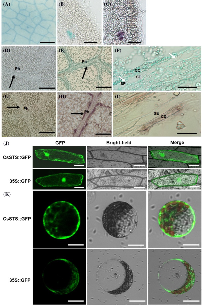
Fig. 2.
Histochemical and subcellular localization of CsSTS. Histochemical analysis of pCsSTS::GUS plants (a, b, d, e, f); immunolocalization of CsSTS in cucumber leaves (c, g, h, i). The section of the leaf without GUS expression (d); The control sections treated with pre-immune serum (g). Longitudinal sections (d–i); transverse sections (b, c). Black arrows indicate the Ph in (d, e, g,h); white arrows in f indicate the SP. Subcellular localization of CsSTS protein in onion epidermal cells (j); Subcellular localization of CsSTS::GFP fusion protein in cucumber mesophyll protoplasts (k). CC companion cells, SP sieve plate, SE sieve element, Ph phloem. Scale bars denote: a 2 mm, b and c 100 μm; d, e, g, h 200 μm; f, i, j 50 μm; k 20 μm

