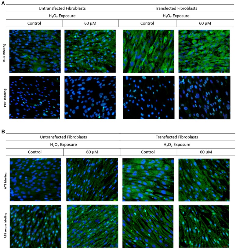
Figure 4.
Behavior of tau in fibroblasts upon oxidative stress and overexpression. Fibroblasts cultures were subjected to transfection with the tau gene and exposed to H2O2 to compare the behavior of Tau to that of untrasfected fibroblasts (UnFs). (A) Tau5 labeling revealed increased cytoplasmic reactivity in TFs exposed to 20 and 60 μM H2O2. PHF reactivity in UnFs was located into the nucleus and is increased in TFs. (B) AT8 labeling of UnFs revealed high cytoplasmic reactivity, which increased in TFs and appears into the nucleus. Anti-AT8 serum showed a similar pattern of that of the AT8 antibody but with a higher signal. Scale bar 2,000 pixels, equivalent to 100 μm. Nuclei were stained with DAPI (in blue), while Tau was detected through FITC conjugated to the secondary antibody (in green). DAPI/FITC signal overlap is observed in green/white.

