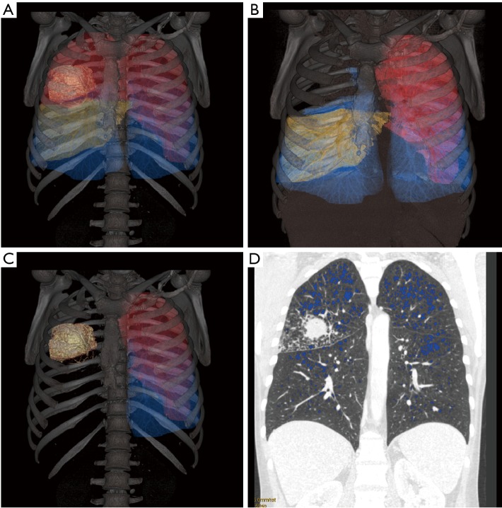
Figure 1.
Quantitative CT of a patient with right upper lobe tumor. (A) Standard 3D reconstruction; (B) 3D reconstruction excluding the right upper lobe; (C) 3D reconstruction excluding the right lung; (D) coronal CT scan showing emphysema lesions (blue) defined by a −950 HU limit and right upper lobe tumor. CT, computed tomography.

