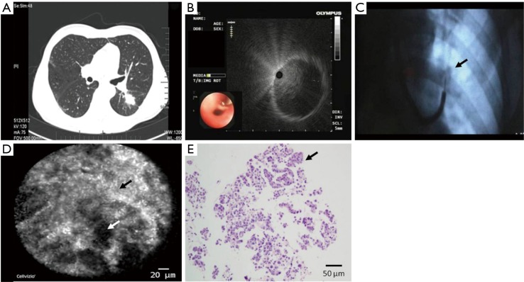
Figure 1.
Images of peripheral pulmonary nodule obtained from CT, EBUS, X-ray, nCLE and histopathologic examination in case 1. (A) The chest CT showed soft-tissue mass in the dorsal segment of left lower lobe, with no bronchial sign; (B) radial probe endobronchial ultrasound confirmed the lesion location outside the bronchial lumen; (C) the puncture needle was inserted into the lesion (black arrow) under the guidance of C-arm X-ray; (D) the confocal endomicroscopic images showed highlighted fibrous tissues (black arrow) with a disordered arrangement, accompanied with spot or black hole structures (white arrow); (E) the histopathological images showed acinous, papillary cancer cells (black arrow) in the lung tissues (hematoxylin-eosin stain, 200×). nCLE, novel needle-based confocal laser endomicroscopy.

