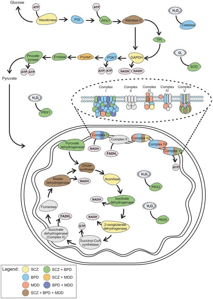
Figure 2.
Schematic representation of the altered proteins in SCZ, BPD, and MDD. Color from each disorder and combination of disorders correlates with the Venn diagram (Figure 1). GAPDH, Glyceraldehyde 3-phosphate dehydrogenase; PFK1, Phosphofructokinase 1; PGAM, Phosphoglycerate mutase; PGI, Phosphoglucose isomerase; PGK1, Phosphoglycerate kinase 1; PRX, Peroxiredoxin; SOD, Superoxide dismutase; TPI, Triosephosphate isomerase.

