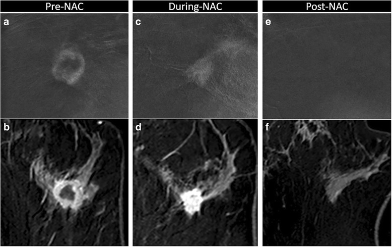
Fig. 6.

Variation of enhancement during neoadjuvant chemotherapy (NAC). Complete response to NAC in a 41-year-old woman with 27 mm invasive ductal carcinoma (G3, triple negative) in the left breast, correctly assessed with contrast-enhanced spectral mammography (CESM) (a, b, c magnification of craniocaudal recombined image) and magnetic resonance imaging (MRI) (b, d, f axial post-contrast T1-w eighted at peak enhancement). Pre-NAC, evaluation showed the same round shape, well-defined margins and rim enhancement on CESM (a) and on MRI (b). During-NAC, both on CESM (c) and on MRI (d) we noted concentric shrinkage, and the tumor was no longer rimmed, with no significant loss of enhancement intensity. Post-NAC, no residual pathological enhancement was visible on CESM (e) or on MRI (f). Complete response was confirmed by the histopathological examination of the surgical specimen
