Fig. 5.
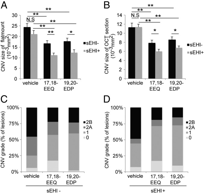
sEH inhibition enhances the protective effect of CYP-derived ω-3 fatty acids on CNV. (A and B) Lesion size at 7 d after CNV induction was assessed by staining of choroidal flat-mount preparations with fluorescent isolectin B4 (A), and a cross-sectional area of lesions was quantified by SD-OCT (B), for C57BL/6 mice administered control vehicle, 17,18-EEQ (50 μg/kg/d), or 19,20-EDP (50 μg/kg/d) once a day without sEH inhibitor (black bars) or with sEH inhibitor (1 mg/kg/d, gray bars). Mice were fed a control diet over the course of the experiment. Data are presented as means ± SEM. *P < 0.05, **P < 0.01. n = 35–43 lesions per experimental group. (C and D) Fluorescein leakage in CNV lesions was graded at 7 d after CNV induction in C57BL/6 mice administered control vehicle, 17,18-EEQ (50 μg/kg/d), or 19,20-EDP (50 μg/kg/d) once a day without sEH inhibitor (C) or with sEH inhibitor (1 mg/kg/d) (D). The grade of the hyperfluorescent lesions is as follows: score 0 (i.e., no leakage); score 1 (i.e., debatable leakage); score 2A (i.e., definite leakage); score 2B (i.e., clinically significant leakage). Mice were fed a control diet over the course of the experiment. n = 35–43 lesions per experimental group.

