Fig. 2.
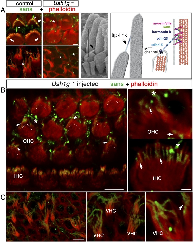
AAV8-Sans-IRES-GFP restores sans expression and targeting in the inner ear hair cells of Ush1g−/− mice. (A, Left) OHC (Upper) and VHC (Lower) hair bundles from P8.5 wild-type (control) and Ush1g−/− mice, immunostained for sans (green) and stained for F-actin with phalloidin (red). Sans is detected at the tips of the stereocilia in the wild-type mouse (white arrowheads), but not in the Ush1g−/− mouse. A nonspecific staining of the kinocilium is present both in the wild-type and Ush1g−/− mice (asterisks). (A, Center) Scanning electron micrograph of the IHC hair bundle, showing the tip links between adjacent stereocilia of different rows (black arrowhead). (A, Right) Diagram showing the tip-link lower and upper insertion points, the position of the mechanoelectrical transduction (MET) channel(s) at the tip of the shorter stereocilium, and the locations of the five USH1 proteins forming the tip link [cadherin-related proteins 15 (cdhr15) and 23 (cdhr23)] or presumably involved in its anchoring to the actin filaments of the taller stereocilium (harmonin b, sans, and myosin VIIa). The submembrane scaffold protein sans belongs to the tip-link upper insertion point molecular complex. Top views of the organ of Corti in the cochlear apical region (B) and of the utricular macula (C) of an injected Ush1g−/− mouse on P8.5 and high-magnification photographs of OHC, IHC, and VHC hair bundles are shown. Sans is targeted to the tips of the stereocilia in all hair cell types (arrowheads). The image in B is extracted from a larger tile scan and contains two tiles stitched together at the upper part of the image. Dashed lines in B indicate the position of the hair bundle base (V shape) in OHCs expressing the transgene. (Scale bars: 5 μm.)

