Fig. 3.
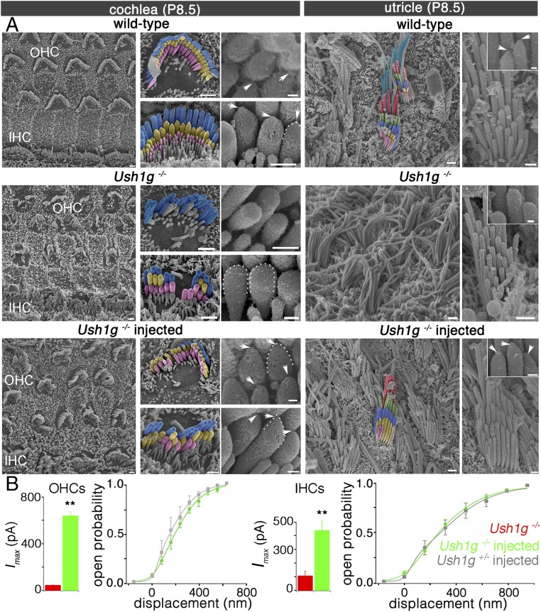
Sans cDNA transfer to inner ear hair cells rescues Ush1g−/− mice from hair bundle structural defects. (A) Low-, intermediate-, and high-magnification scanning electron micrographs showing the architecture of the hair bundles in cochlear OHCs and IHCs (Left) and in VHCs (utricle, Right) of a control wild-type mouse, an Ush1g−/− mouse, and an Ush1g−/− mouse injected with the Sans cDNA, on P8.5. In the wild-type mouse, the stereocilia tips have prolate shapes, which is the hallmark of functional tip links (arrowheads and dashed lines) (31), whereas they have rounded shapes in the Ush1g−/− mouse (dashed lines), in keeping with the loss of the tip links (24). Note the fragmentation of the hair bundles and the degeneration of some stereocilia in this mouse. In the injected Ush1g−/− mouse, the hair bundles have recovered their normal staircase architecture and the prolate shapes of stereocilia tips (arrowheads and dashed line). (Scale bars: 2 μm.) (B) Mechanoelectrical transduction (MET) currents recorded ex vivo in IHCs and OHCs of uninjected Ush1g−/− (red), injected Ush1g−/− (green), and injected Ush1g+/− (control, gray) P8.5 mice. Bar charts show the peak amplitudes of the MET currents (Imax) recorded in the hair cells of uninjected or injected Ush1g−/− mice (110.8 ± 30.8 pA in IHCs and 47.3 ± 5.7 pA in OHCs of uninjected Ush1g−/− mice vs. 424 ± 70 pA in IHCs and 641 ± 35 pA in OHCs of injected Ush1g−/− mice). Note the marked increase of Imax in the injected Ush1g−/− mice compared with the noninjected mice (Student’s t test, **P < 0.01 for both IHCs and OHCs). Line graphs show the MET channel opening probability plotted as a function of hair bundle displacement for IHCs and OHCs of injected Ush1g+/− (gray) and Ush1g−/− (green) mice. For both cell types, the two curves are superimposed.

