Fig. S1.
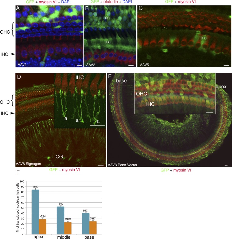
Cell tropism of AAV1, AAV2, AAV5, and AAV8 in the cochlea. The recombinant vectors AAV1 and AAV8 from SignaGen Laboratories or AAV2, AAV5, and AAV8 from Penn Vector Core, all containing the GFP reporter gene, were injected on P2.5 through the round window membrane into the left cochlea of 9, 6, 8, 8, and 31 mice, respectively. The sensory epithelium of the cochlea (organ of Corti) was microdissected on P8.5, and immunolabeled for myosin VI or for otoferlin, to visualize all hair cells or only IHCs, respectively, and for GFP. (A and B) Cell nuclei were stained blue with DAPI. (A–C) AAV1 (SignaGen Laboratories) and AAV2 (Penn Vector Core) mostly transduce supporting cells, namely, inner phalangeal cells (IPC) and/or Deiters’ cells (DC), and AAV5 transduces only a few supporting cells. AAV8-CAG from SignaGen Laboratories mostly transduces the cochlear ganglion (CG) neurons but not the hair cells (D) [Inset, detailed view of the IHCs and afferent nerve terminals (a) is shown], whereas AAV8-CAG from Penn Vector Core transduces both IHCs and OHCs (E), more efficiently in the apical region (E, Inset) than in the basal region of the cochlea as shown in a tile scan of images stitched into a large mosaic of nearly the entire organ of Corti (E). (F) Bar chart showing the proportions of IHCs and OHCs transduced with AAV8-CAG from Penn Vector Core in the apical, middle, and basal regions of the cochlea. (Scale bars: A–C, 5 μm; D and E, Inset, 10 μm; E, 50 μm).

