Fig. 5.
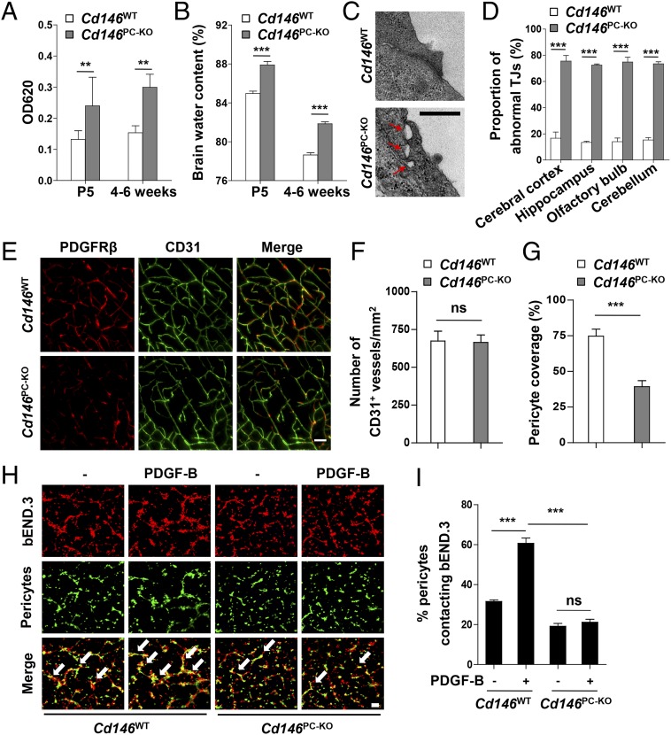
Pericyte Cd146 deletion results in impaired pericyte recruitment and BBB breakdown. (A) Cd146WT and Cd146PC-KO mice at P5 or 4–6 wk were given an i.p. injection or i.v. injection of Evans blue dye, respectively, and the absorption of Evans blue extracted from the mouse brain was measured by a microplate spectrophotometer at 620 nm. (B) Brain water content of Cd146WT and Cd146PC-KO mice (P5 or 4–6 wk). (C) EM images of TJs of brain capillaries in the cortex from Cd146WT and Cd146PC-KO mice (P5). Red arrows indicate altered TJ alignment. (Scale bar: 500 nm.) (D) Quantification of the abnormal TJ structure of brain capillaries in cerebral cortex, hippocampus, olfactory bulb, and cerebellum from Cd146WT and Cd146PC-KO mice (P5; at least 50 TJs were analyzed per group). (E) Brain sections from the cortex of mice at P5 were costained for CD31 (green) and PDGFRβ (red) and analyzed by LSFM after being optically cleared by using organic solvents. Representative MIPs of 40 virtual single slices from Cd146WT and Cd146PC-KO mice are shown. (Scale bar: 50 μm.) (F) Quantification of the number of CD31+ capillaries in cortex from Cd146WT and Cd146PC-KO mice. No difference was detected. (G) Pericyte coverage was quantified by analyzing percent length of CD31+ capillaries opposed to PDGFRβ+ pericytes. Decreased pericyte coverage in capillaries was observed in the cortex of Cd146PC-KO mice. (H) bEND.3 cells and brain microvessel pericytes isolated from Cd146WT and Cd146PC-KO mice were labeled by PKH26-red and CFSE, respectively, and were cocultured in Matrigel-coated culture slides for 6 h. White arrows indicate pericytes contacting bEND.3 cells. (Scale bar: 100 μm.) (I) Quantification was performed by measuring the merged cells from 15 fields of 3 independent experiments (**P < 0.01 and ***P < 0.001). Data are from one experiment representative of three independent experiments with eight mice per genotype (A–D) or five mice per genotype, at least eight MIPs per mouse, and five random fields per MIP (F and G).

