Fig. 6.
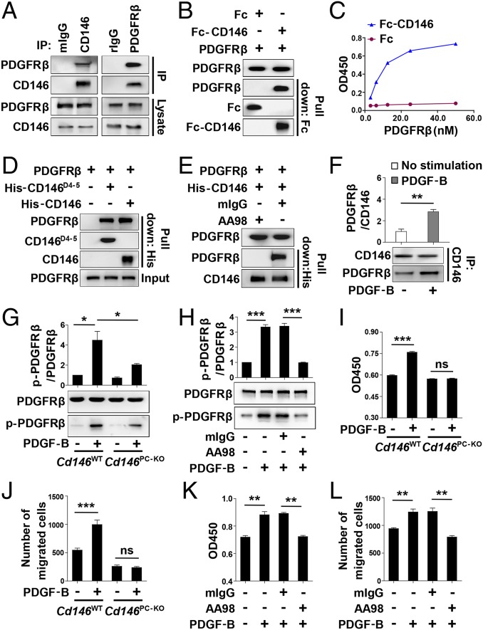
CD146 activates PDGFRβ and promotes pericyte proliferation and migration. (A) Coimmunoprecipitation of endogenous CD146 and PDGFRβ in 10T1/2 cells. (B) Direct interaction between CD146 and PDGFRβ detected by Fc pull-down assay. (C) Fc or Fc-CD146 was added to wells coated with different concentrations of PDGFRβ, and ELISA was performed. (D) PDGFRβ directly binds CD146D4-5. PDGFRβ was first incubated with His-tagged CD146-ECD or CD146D4-5, respectively. The complex was pulled down by anti-His antibody. (E) Anti-CD146 AA98 abrogates CD146 and PDGFRβ interaction. His-CD146-ECD was incubated with PDGFRβ in the presence of mIgG or AA98 (50 μg/mL). (F) PDGF-B stimulation enhances the interaction of CD146 and PDGFRβ. Pericytes were stimulated without or with PDGF-B (20 ng/mL). (G) Cd146 deletion leads to impaired PDGFRβ activation. CNS pericytes isolated from Cd146WT and Cd146PC-KO mice were incubated with PDGF-B (20 ng/mL). The level of p-PDGFRβ was detected by Western blotting. (H) AA98 impairs PDGF-B–induced PDGFRβ activation in CNS pericytes isolated from Cd146WT mice. (I and J) The proliferation and migration of CNS pericytes isolated from Cd146WT and Cd146PC-KO mice in the presence of PDGF-B (20 ng/mL) were determined by CCK-8 assay (I) and transwell Boyden chamber assay (J). (K and L) Proliferation and migration of CNS pericytes from Cd146WT mice during PDGF-B (20 ng/mL) stimulation without or with anti-CD146 AA98 (50 μg/mL) were determined by CCK-8 assay (K) and transwell Boyden chamber assay (L). Data represent three independent experiments (*P < 0.05, **P < 0.01, and ***P < 0.001).

