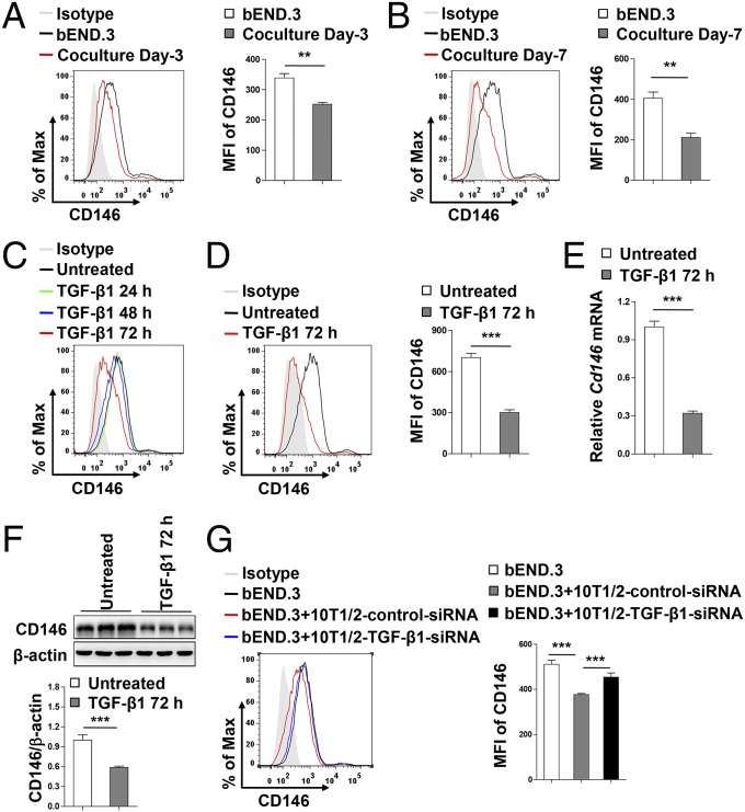
Fig. 7.
Pericytes down-regulate endothelial CD146 expression through TGF-β1. (A and B) Flow-cytometry analysis of CD146 expression in bEND.3 cells cocultured without or with pericytes for 3 d (A) or 7 d (B). (C) Flow-cytometry analysis of CD146 expression in bEND.3 cells stimulated without or with TGF-β1 (10 ng/mL) for 24 h, 48 h, and 72 h. (D–F) bEND.3 cells were stimulated without or with TGF-β1 (10 ng/mL) for 72 h. Expression of CD146 in bEND.3 was determined by flow cytometry (D), real-time PCR (E), or Western blotting (F). (G) 10T1/2 cells transfected with control siRNA or TGF-β1 siRNA were cocultured with bEND.3 cells. The expression of CD146 in bEND.3 was determined by flow cytometry. (Right) Quantification of the MFI of CD146 (**P < 0.01 and ***P < 0.001). Data represent three independent experiments.

