Fig. S1.
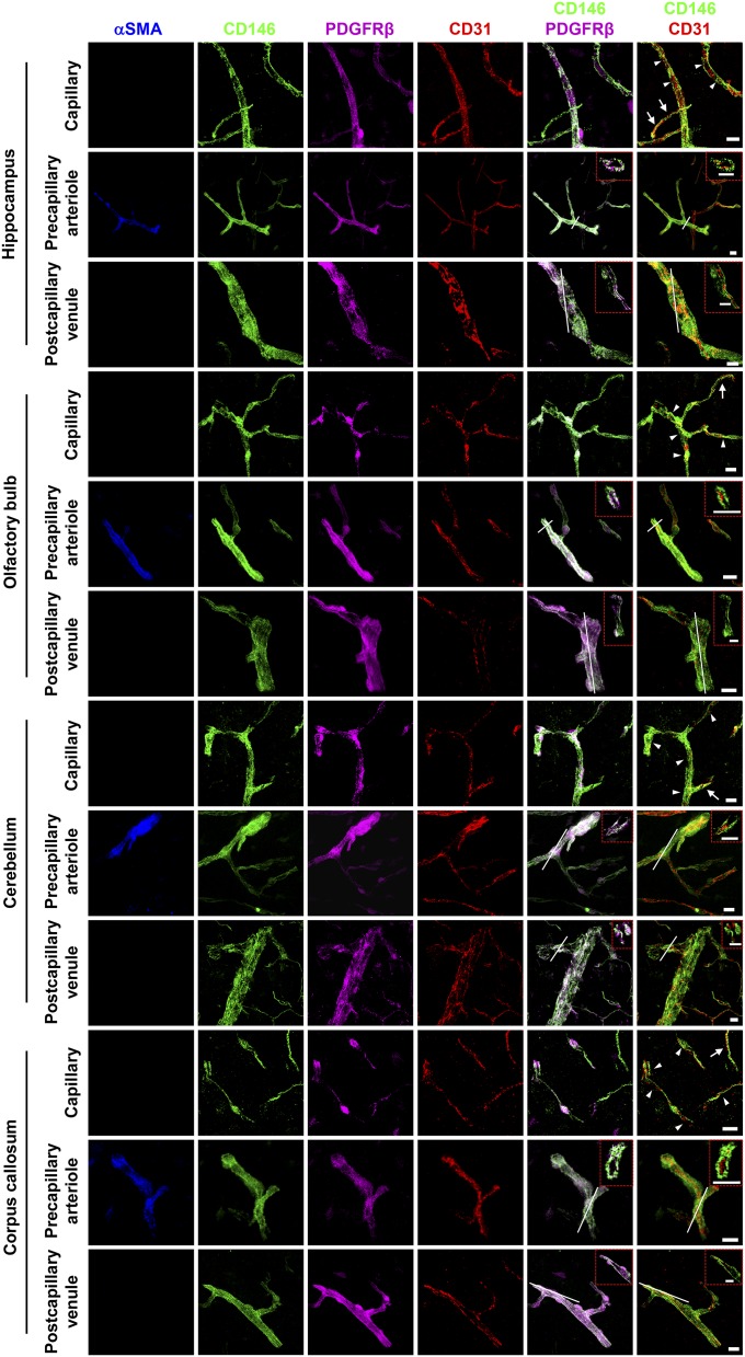
The expression of CD146 in brain vessels at 4–6 wk. Brain sections (40-μm thickness) from the hippocampus, olfactory bulb, cerebellum, and corpus callosum (white matter) of mice at 4–6 wk were stained for CD146 (green), CD31 (EC marker; red), PDGFRβ (pericyte marker; purple), and αSMA (artery marker; blue). Shown are 3D reconstructions of confocal image z-stacks of brain capillaries, precapillary arterioles, and postcapillary venules. The same expression pattern of CD146 was observed in different brain regions where CD146 was expressed in the BECs of immature capillaries without pericyte coverage (arrows) and was only expressed in pericytes of brain capillaries covered with pericytes (arrowheads), precapillary arterioles, and postcapillary venules. The dashed red rectangles indicate a confocal z-slice located at the white line of each panel, confirming that the expression of CD146 was exclusively observed in pericytes but not in BECs of precapillary arterioles and postcapillary venules. At least 20 capillaries, 20 precapillary arterioles, and 20 postcapillary venules from the hippocampus, olfactory bulb, cerebellum, and corpus callosum were analyzed. (Scale bars: 10 μm.) Data are from one experiment representative of three independent experiments with five mice.

