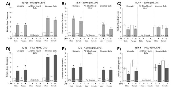
Figure 1. Experiment 2: Relative Gene Expression of IL-1β, IL-6, and TLR-4.
A and D) Relative gene expression of IL-1β significantly increased in microglia treated with LPS compared to vehicle-treated microglia. IL-1β expression was not detected in the microglia-depleted/all other neural cell population. IL-1β expression significantly increased in unsorted cells treated with LPS compared to vehicle-treated unsorted cells. B) Relative gene expression of IL-6 significantly increased in microglia treated with a 500 ng/mL LPS dose compared to vehicle-treated microglia. IL-6 expression was not detected in the microglia-depleted/all other neural cell population. IL-6 expression significantly increased in unsorted cells treated with a 500 ng/mL LPS dose compared to vehicle-treated unsorted cells; and, the LPS-facilitated increase in IL-6 expression was greater in the male unsorted cells compared to the female unsorted cells. C and F) Relative gene expression of TLR-4 significantly decreased in microglia treated with LPS compared to vehicle-treated microglia. TLR-4 expression was not detected in the microglia-depleted/all other neural cell population. No significant differences were found in TLR-4 expression in the unsorted cells. E) Relative gene expression of IL-6 significantly increased in microglia treated with a 1,000 ng/mL LPS dose compared to vehicle-treated microglia. IL-6 expression was not detected in the microglia-depleted/all other neural cell population. IL-6 expression significantly increased in unsorted cells treated with a 1,000 ng/mL LPS dose compared to vehicle-treated unsorted cells. *p < 0.05

