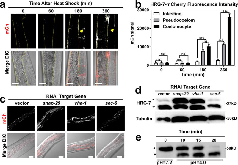
Figure 3. HRG-7 secretion and maturation is regulated by specific trafficking factors.

a) mCherry expression in strain IQ7170 (Phsp-16.2::HRG-7::mCherry) placed in 37°C for 30 minutes, then 20°C for 60, 180, and 360 minutes. Dotted yellow lines outline the intestine. Arrowheads indicate HRG-7::mCherry in the pseudocoelom. Scale bar = 20 μm. Images are representative of three independent experiments. b) Quantification of mCherry in coelomocytes, pseudocoelom, and intestine of IQ7170 following heat shock. For quantification, 10 images, consisting of 10-15 Z stacks per image, were used for each timepoint. ***P<0.001, **P<0.01, *P<0.05 (two-way ANOVA). mCherry signal is in arbitrary unites. Graph and statistics are from a single experiment. The experiment was repeated one time with similar results. c) mCherry expression in strain IQ7670 fed dsRNA against vector, snap-29, vha-1, and sec-6. The small puncta visible in all panels are autofluorescent gut granules in the intestine. Scale bar = 20 μm. Images are representative of three independent experiments. d) Immunoblot analysis of strain N2 fed dsRNA against vector, snap-29, vha-1, and sec-6. Membranes were probed with polyclonal anti-HRG-7 antibody and then incubated with HRP-conjugated anti-rabbit secondary antibody. * indicates pro-HRG-7. + indicates mature HRG-7. Unprocessed blots are shown in Figure S6. Data are representative of three independent experiments. e) Immunoblot analysis of lysate prepared from strain IQ7370 (Pvha-6::HRG-7::3xFLAG) at pH 7.2 and pH 4. Membranes were probed with monoclonal anti-FLAG antibody and then incubated with HRP-conjugated anti-mouse secondary antibody. * indicates pro-HRG-7-3xFLAG. + indicates mature HRG-7-3xFLAG. Unprocessed blots are shown in Figure S6. Data are representative of three independent experiments.
