Figure 3.

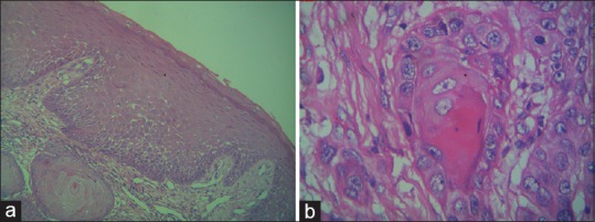
(a) Photomicrograph showing features of well-differentiated squamous cell carcinoma – low magnification (×10). (b) Photomicrograph showing features of well-differentiated squamous cell carcinoma – high magnification (×40)

(a) Photomicrograph showing features of well-differentiated squamous cell carcinoma – low magnification (×10). (b) Photomicrograph showing features of well-differentiated squamous cell carcinoma – high magnification (×40)