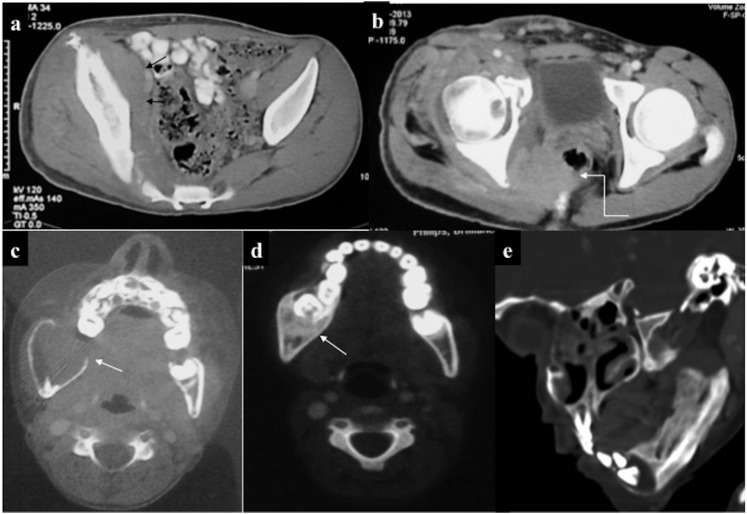
Figure 19.
Musculoskeletal chloroma: (a, b) contrast-enhanced CT of the pelvis of a leukaemic patient revealed ill-defined isodense soft tissue in the right hemipelvis (arrow) infiltrating the iliopsoas and piriformis muscles. Furthermore, it was infiltrating the mesorectal fascia with rectal involvement (curved arrow). Additionally, there was mild expansion and cortical thickening of the right iliac bone. (c, d) A different case with lytic expansile lesion with cortical thinning involving the ramus of the right hemimandible: biopsy revealed chloroma. 8 months post-chemotherapy, lesion had become sclerotic with no soft tissue component. (e) Another patient showing predominantly sclerotic involvement of the left hemimandible.

