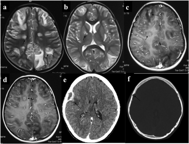
Figure 3.
Intra-axial central nervous system chloroma (a–d): a 5-year-old male with acute myeloid leukaemia presented with features of raised intracranial tension and generalized tonic–clonic seizures. MRI showed multiple circumscribed lesions of intermediate T2 signal intensity (a, b) involving bilateral cerebral hemispheres including basal ganglia with perilesional oedema (PLE). On contrast-enhanced MRI, these lesions showed solid or ring enhancement. Additionally, there was patchy leptomeningeal enhancement (arrows in c). (e, f) CECT of a 26-year-old leukaemic patient performed for headache showed lenticular extra-axial mass with peripheral enhancement and focal bony erosion in the right frontal lobe. Medial border of this lesion was irregular with nodular component showing parenchymal invasion and PLE.

