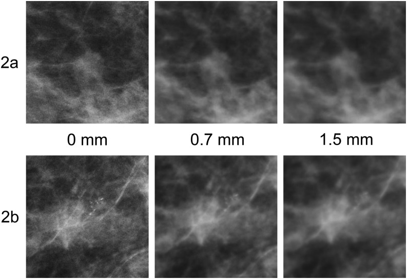
Figure 2.
(a, b) Zoomed areas of full-field digital mammography images at 0, 0.7 and 1.5 mm of simulated blurring. (a) A spiculated mass of irregular shape with indefinite borders. (b) A single cluster of granular microcalcifications with different shapes, densities and sizes. Although the mass becomes increasingly difficult to visualize, the microcalcifications are no longer visible with 1.5 mm of simulated blur.

