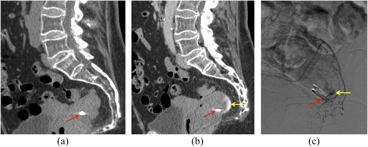
Figure 7.
In a 72-year-old female with gastrointestinal bleeding following rectal biopsy, the clip (arrows) can be seen on non-contrast image (a), with active contrast extravasation present in the rectum on portal venous phase image (b, arrow). Catheter angiogram of a rectal branch (c) confirms a site of active extravasation (arrow) adjacent to the rectal clip. Embolization was performed subsequently.

