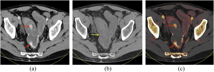
Figure 8.
A 51-year-old male with prior colectomy for clostridium difficile colitis presenting with rectal bleeding: “mixed” image from a dual-energy CT scan in the portal venous phase (a) shows hyperattenuating material within the distal small bowel and rectum (arrows). On the virtual non-contrast (VNC) image (b), this material is not present within the bowel lumen, showing that this is not ingested material. The anastomotic suture material remains visible on the VNC image (arrow). On iodine overlay image (c) with iodine content colour-coded in orange, the hyperattenuating material within the bowel lumen demonstrates iodine content, further confirming that this represents active extravasation.

