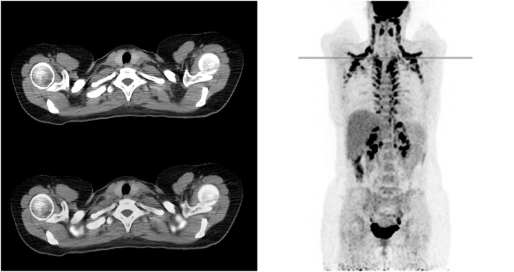
Figure 1.
An example of a fluorine-18 fludeoxyglucose positron emission tomography/CT scan of a patient with high brown adipose tissue (BAT) uptake in the neck, interscapular, paravertebral and perinephric regions is shown. BAT can usually be distinguished from pathological uptake by its typical pattern and low attenuation in the CT scans.

