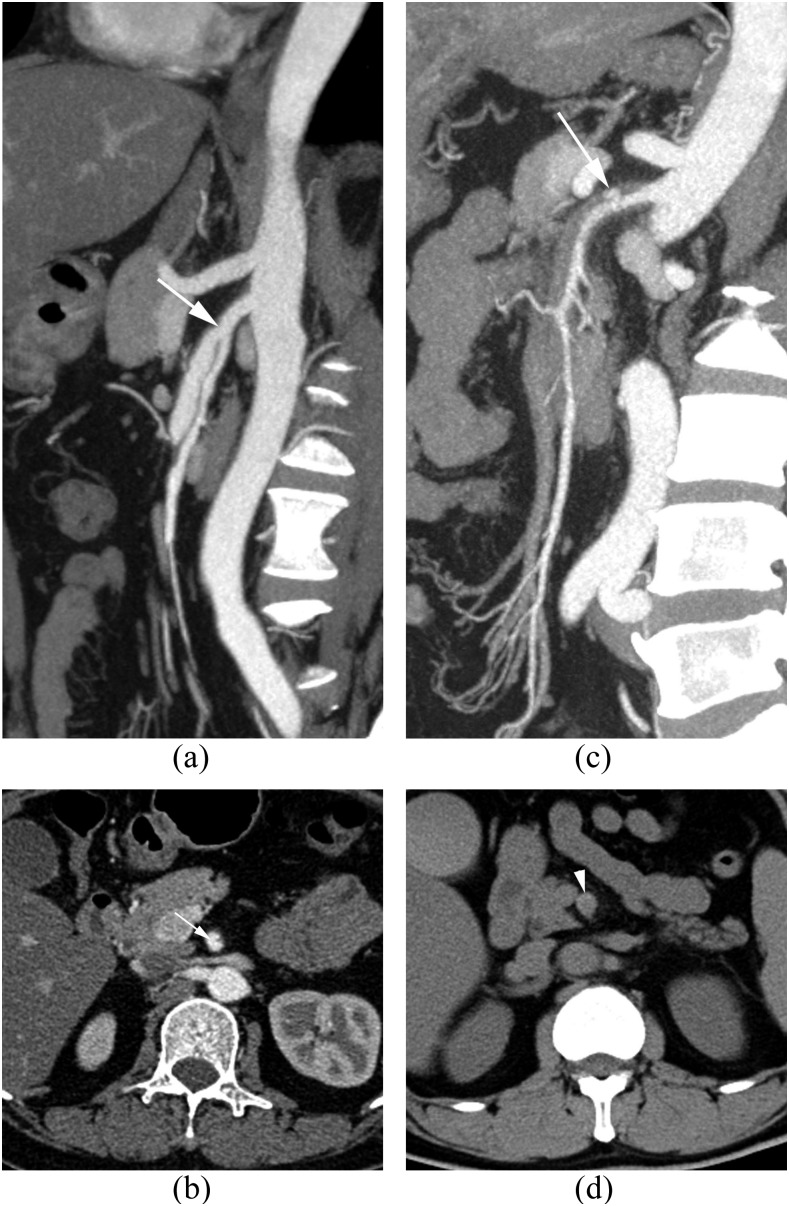
Figure 2.
A 47-year-old female patient with Type IIa spontaneous isolated dissection (SID) of the superior mesenteric artery (SMA) (a, b) and a 48-year-old male patient with Type IIb SIDSMA (c, d). Sagittal and transverse views of post-contrast-enhanced images show the entry site (white arrows) of the dissection (a–c) located in the anterior wall of the SMA; the transverse view before contrast enhancement shows the crescent-shaped high attenuation (white arrowhead) in the SMA (d).

