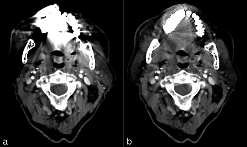
Figure 4.
Images from the CT examination of a 72-year-old male after neck dissection and resection of a squamous cell carcinoma of the maxilla. CT slice at the level of the oropharynx before (a) and after (b) reconstruction with iterative metal artefact reduction (iMAR). The image before iMAR was severely distorted with artefacts owing to metal hardware in the maxilla, which affected the visualization of the anatomy, especially in the regions next to the metal implants. After the application of iMAR, a considerable reduction of artefacts and improvement in image quality was observed with visualization of the anatomy, especially next to the metal hardware. It can be noted that there is no change of quality in the regions less affected by artefacts, for example paraspinal regions.

