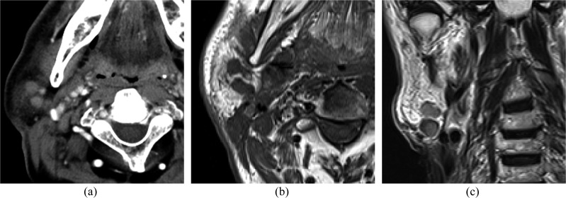
Figure 3.
A 70-year-old male with metastases to the parotid nodes from squamous cell carcinoma of the larynx: (a) an axial post-contrast CT image is showing multiple homogeneous enhancing masses in the parotid tail. (b, c) Axial T1 weighted (b) and coronal T2 weighted (c) images are showing multiple well-defined masses in the parotid tail.

