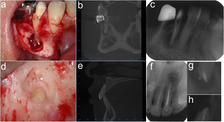
Figure 1.
Apical extension of root canal obturation: Tooth 44—obturation was determined as “overextension” by photomicrography (a) and classified as “overextension” by CBCT (b), while as “no overextension” by periapical radiography (PR) (c). Tooth 21: obturation was determined as “overextension” by microphotography (d) and classified as “no overextension” by CBCT (e) and PR (f). Apical areas of the CBCT images (b, e) were magnified (g, h) respectively.

