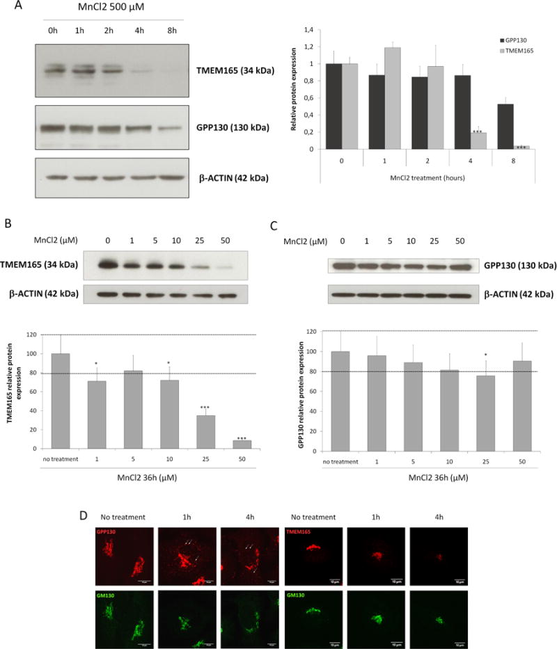
Figure 1. TMEM is rapidly degraded in response to Mn.

(A) Steady state cellular level of TMEM165 and GPP130. HeLa cells were treated with MnCl2 500 μM for 0 to 8h. Total cell lysates were prepared, subjected to SDS-PAGE and Western blot with the indicated antibodies. Right panel shows the quantification of TMEM165 and GPP130 protein levels after normalization with actin (Number of experiments (N) = 2; *** = P value < 0,001). (B) Steady state cellular level of TMEM165. HEK293 cells were treated with MnCl2 from 0 to 50 μM for 36h. Total cell lysates were prepared, subjected to SDS-PAGE and Western blot with the indicated antibodies. Lower panel shows the quantification of TMEM165 protein levels after normalization with actin (N = 2; *** = P value < 0,001). (C) Steady state cellular level of GPP130 in the same experimental conditions as described in (B). (D) HeLa cells were incubated with MnCl2 500 μM for 1 and 4h, fixed and labeled with antibodies against TMEM165, GPP130 and GM130 before confocal microscopy visualization. White arrows point to some GPP130 positive vesicles.
