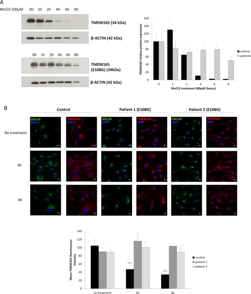
Figure 6. The ELDGK motif is crucial for Mn2+ sensitivity.

(A) Healthy skin fibroblasts (upper left) and patients skin fibroblasts (lower left) carrying E108G mutation were treated with MnCl2 500μM for 0 to 8h. Total cell lysates were prepared, subjected to SDS-PAGE and Western blot with the indicated antibodies. Right panel shows the quantification of TMEM165 protein levels after normalization with actin (N = 2; *** = P value < 0,001). (B) Healthy skin fibroblasts and patients skin fibroblasts carrying E108G mutation were treated with MnCl2 500μM for 0, 4 and 8h. Cells were then fixed and labeled with antibodies against TMEM165 and GM130 before confocal microscopy visualization (N = 2; *** = P value < 0,001). Lower panel shows the quantification of the associated TMEM165 fluorescence intensity (N = 2; n = 30; *** = P value < 0,001).
