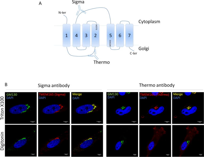
Figure 8. TMEM165 topology.

(A) Representation of TMEM165 predicted topology. The two anti-TMEM165 antibodies (Sigma-Aldrich and Thermo Fisher Scientific) depicted here recognize two different parts of the protein. The Sigma one recognize the cytoplasmic loop between the fourth and fifth transmembrane domains. The Thermo one recognize the short luminal loop between the first and the second transmembrane domain. (B) Cells were fixed with paraformaldehyde 4% and treated as described in materials and methods. Selective permeabilization was done by using triton ×100 or digitonin. Cells were labeled with antibodies against TMEM165 and GM130 before confocal microscopy visualization.
