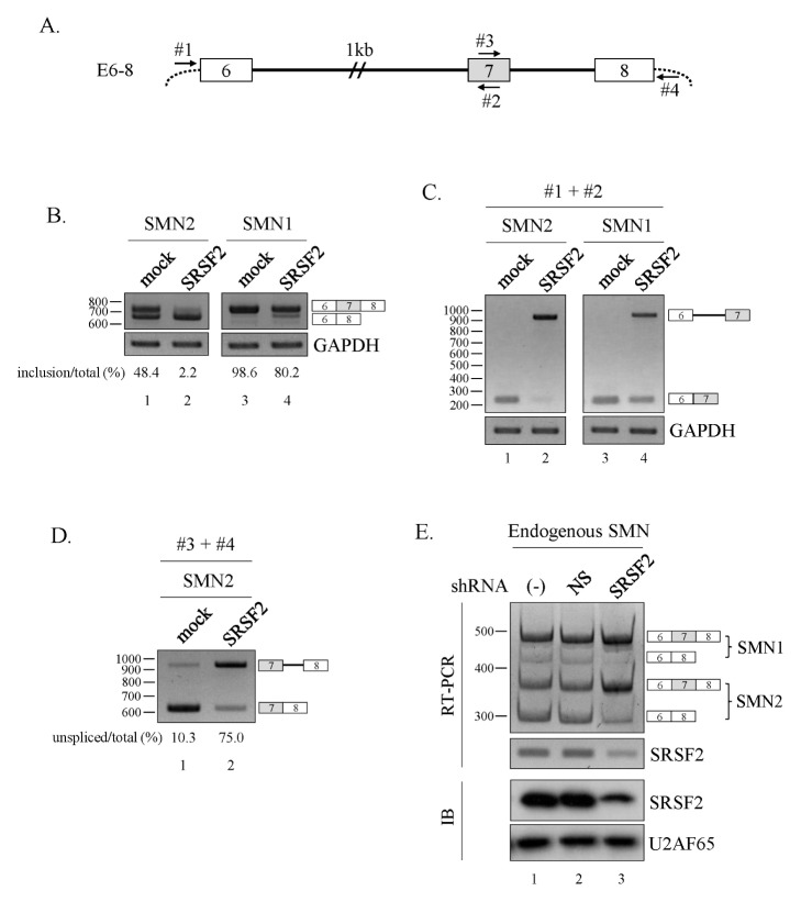
Fig. 1.
SRSF2 promotes exon exclusion. (A) Schematic diagram of the E6–8 minigene. Exons are depicted as numbered boxes, introns as solid lines. (B) RT-PCR analysis of the E6–8 minigene from the SMN1/2 locus in SRSF2-expressing cells. (C) RT-PCR analysis of intron6 splicing within the E6–8 minigene using primers #1 and #2. (D) RT-PCR analysis of intron7 splicing within the E6–8 minigene using primers #3 and #4. (E) RT-PCR analysis to detect alternative splicing of endogenous SMN1 and SMN2 using RNA extracted from cells infected with lentiviruses with SRSF2-targeting shRNA (SRSF2) or non-silencing shRNAs (NS).

