
Figure 3. Detailed Interactions of AcrIIA4 with sgRNA-bound SpyCas9.
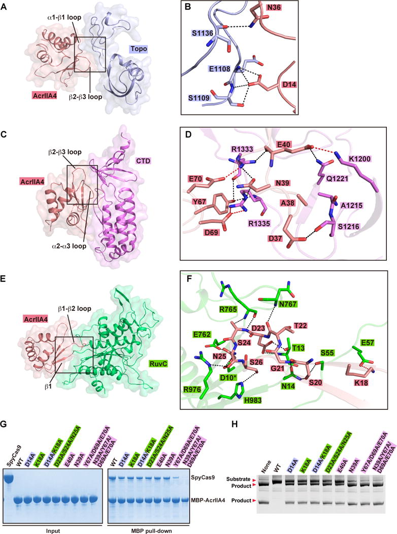
(A, C, E) Surface views of the interfaces between AcrIIA4 and Topo domain (panel A), CTD domain (panel C), and RuvC domain (panel E). The interface segments are highlighted by black boxes.
(B, D, F) Detailed interactions at the interfaces between AcrIIA4 and Topo domain (panel B), CTD domain (panel D), and RuvC domain (panel F) are shown in stick representations. The color code is the same as Figure 2A. Hydrogen bonds and salt bridges are colored as black and red dashed lines, respectively.
(G) Mutation analysis of AcrIIA4 residues involving in the binding to sgRNA-bound SpyCas9 by MBP pull-down assay of MBP-tagged AcrIIA4.
(H) In vitro enzymatic assay of Ala mutations of AcrIIA4 residues that impaired or abolished binding of AcrIIA4 to sgRNA-bound SpyCas9.
See also Figure S3.