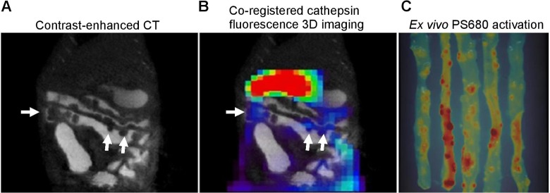
Figure 6.
Dual-model microCT/FMT imaging on intestinal adenomas in Apc Min/+ mice. A, Tumor nodules were detected by contrast-enhanced microCT imaging (white arrows). B, Coregistration of microCT image with 3-dimensional (3D) fluorescence image activated by ProSense 680 probe. Three-dimensional image was taken at 24 hours after injection of ProSense 680 activatable probe and fused with microCT image for anatomical colocalization. C, Two-dimensional fluorescence reflectance imaging (FRI) images of small intestine. After coregistration of in vivo imaging, the small intestine was dissected and imaged ex vivo by FRI for validation.

