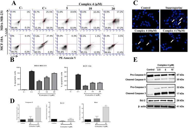
Fig 5. Effect of complex (4) on apoptosis in MDA-MB-231 breast tumor cells and MCF-10A non-tumor breast cells.
(A) After treatment with the indicated concentrations of complex (4), cells were incubated with PE-Annexin-V and 7AAD for 15min, harvested and then analyzed by cytometry. (B) The percentage of apoptotic and necrotic cells was plotted in a graph for MDA-MB-231 and MCF-10A cells. The fluorescence of 7AAD is detected in the FL3-A channel and the fluorescence of PE-Annexin-V is detected in the FL2-A channel. Camptothecin (campto) was used as a positive control for apoptosis. (C) Nuclear fragmentation promoted by complex (4) in MDA-MB-231 cells was investigated using DAPI staining. Staurosporine was used as a positive control for nuclear fragmentation. White arrows show fragmented nuclei. The expression of apoptotic and anti-apoptotic molecules was investigated by (D) qRT-PCR and (E) Western blotting analysis. Significant at the *p<0.005, **p<0.001, ***p<0.0001 levels using ANOVA and Bonferroni tests.

