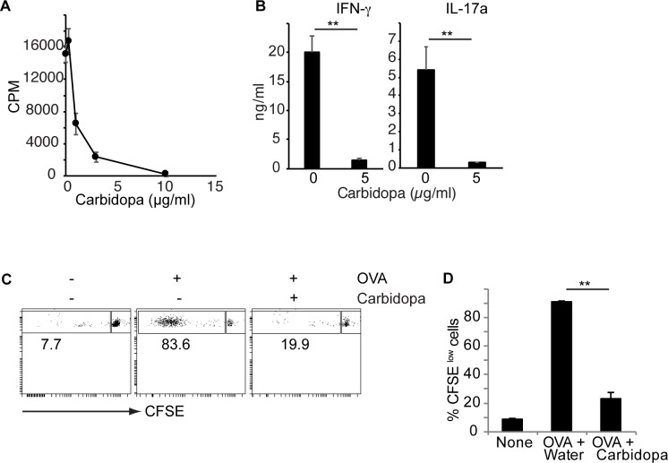
Fig 1. Carbidopa inhibit CD4+ T cell proliferation.
A, CD4+ T cells were culture on anti-CD3 coated plates with or without indicated concentration of carbidopa. Two days later, proliferation of cells was determined by 3H-thymidine assay. Error bars represent standard deviation of triplicates. B, IFN-γ and IL-17a production by anti-CD3 activated naïve CD4+ T cells in the presence or absence of carbidopa. C, OTII CD4+ T cells (Thy1.1+) were transferred into WT C57BL/6 mice (Th1.2+). Next day animals were immunized with OVA. Four days later, CFSE dilution was assessed on Thy1.1+CD4+ T cells. D, Summary of CFSElow cells from data presented in B. (n = 2–3 mice). A representative of 2 experiments is shown. ***P = <0.005, P value <0.05 were considered significant.

