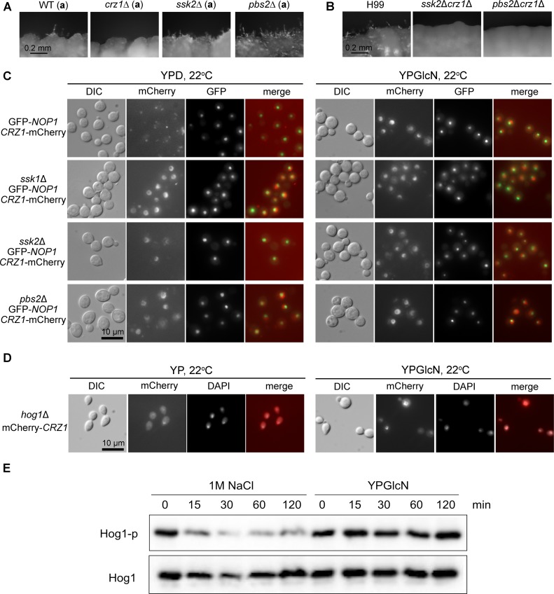
Fig 8. The HOG components function upstream of Crz1 and suppress Crz1’s nuclear localization.
(A) Cells of the wild-type KN99a, crz1Δ (a), ssk2Δ (a), and pbs2Δ (a) were cultured on YP+GlcN (0.5%) medium for 7 days. (B) Cells of the wild-type H99, the ssk2Δcrz1Δ double mutant, and the pbs2Δcrz1Δ double mutant were cultured on YP+GlcN medium for 7 days. (C) The control strain XW252 (GFP-Nop1, PCRZ1-Crz1-mCherry) [79] and the corresponding ssk1Δ, ssk2Δ, and pbs2Δ mutants in the XW252 background were cultured in YPD or YP+GlcN medium overnight at 22°C. GFP-Nop1 labels the nucleolus within the nucleus [79]. (D) The strain JL408 (MATα, hog1Δ; PGPD1-mCherry-CRZ1) was generated from a cross between the hog1Δ mutant in the mating type a background and the strain JL410 (MATα, PGPD1-mCherry-CRZ1). JL408 was cultured in YP or YP+GlcN medium overnight at 22°C. (E) The wild-type strain H99 was grown to mid-logarithmic phase and then exposed to 1 M NaCl or 2% glucosamine (YPGlcN) for the indicated time points. Hog1 phosphorylation levels were monitored using anti-P-p38 antibody. The blot was stripped and then used for detection of Hog1 protein level with a polyclonal anti-Hog1 antibody as a loading control.

