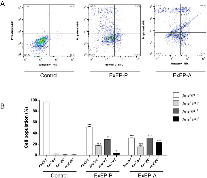
Fig 4. Cell death profile after treatment with IC50 of the ExEP-P and ExEP-A.
(A) Dot plots indicating the flow cytometry, and (B) representative diagrams obtained via flow cytometry of cells stained with annexin V-FITC/PI; Anx–/PI–: viable cells; Anx+/PI–: apoptotic cells; Anx–/PI+: necrotic cells, and Anx+/PI+: cells in late apoptosis. ***p < 0.001 treated group versus control viable cells. ###p < 0.001 treated group versus control apoptosis. +++p < 0.001 treated group versus control necrosis. xxxp < 0.001 and xp < 0.05 treated group versus control late apoptosis.

