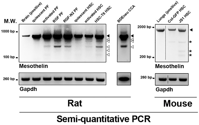
Fig 1. Semi-quantitative RT-PCR of Msln expression in murine liver myofibroblast and cholangiocarcinoma cells.
Expression of Msln transcripts is analyzed in cDNA samples from primary and immortalized mouse and/or rat liver myofibroblasts, and rat cholangiocarcinoma cells. Positive controls include rat brain for rat Msln gene PCR, and mouse lungs for mouse Msln gene. Gapdh is used as reference. Wild-type Msln amplified is observed in all wells for both species (black arrowhead). Rat Msln splicing variants are also observed (empty arrowheads). Reaction artifacts were observed in mouse Msln PCR reactions (asterisks). Primer sequences are listed in Table 1. M.W., molecular weight; bp, base pairs; HSC, hepatic stellate cell; CCA, cholangiocarcinoma; PF, portal fibroblast.

