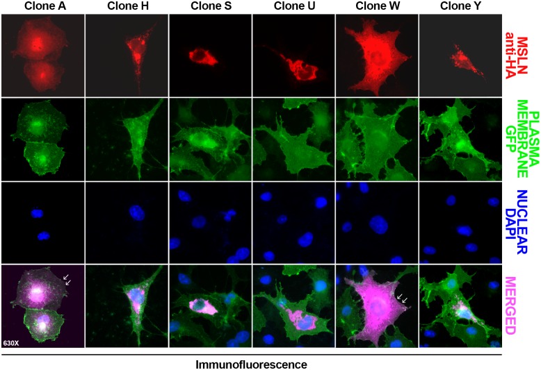
Fig 5. Immunofluorescence analysis of Msln splicing variant distribution in transfected COS7 cells.
COS7 cells transfected with plasmid DNAs encoding Msln splicing variants A, H, S, U, W, and Y (red) were transduced to express plasma membrane-bound GFP (green), and analyzed by microscopy. While recombinant Msln A and W variants are associated with the plasma membrane (white arrowheads) and cytoplasm compartments, the remaining Msln are observed predominantly in the cytoplasmic/perinuclear area of the cells (purple). Nuclear stain is DAPI (blue). Magnification 630X.

