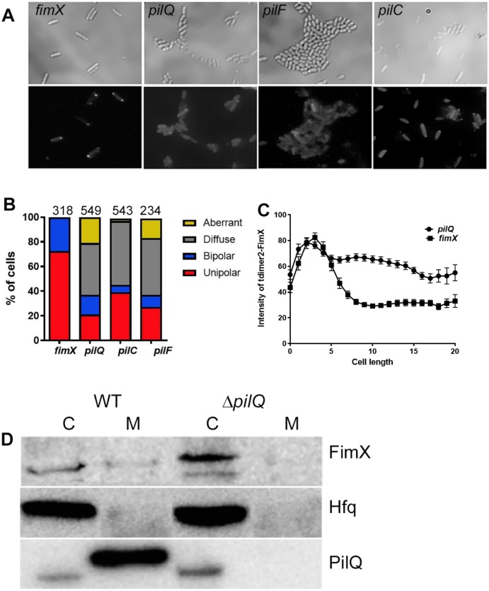
Fig 3. FimX unipolar localization requires structural components of the pilus assembly machinery.
(A) tdimer2-FimX localization in live PA14 fimX, pilQ, pilF or pilC mutants. Plate grown bacteria were spotted onto 1% agar pads. Upper panel, phase contrast; lower panel, tdimer2 epifluorescence. (B) Distribution of tdimer2-FimX in fimX, pilQ, pilF and pilC mutants. The number of cells scored for each strain is noted above each bar. (C) Fluorescence intensity profile of tdimer2-FimX for fimX and pilQ mutants over the length of the cell in pixels (n = 50). (D) Distribution of endogenous FimX between membrane (M) and cytosolic (C) fractions differs in PA14 vs. pilQ::Tn bacteria. Samples were immunoblotted for FimX, the cytosolic protein Hfq, and the outer membrane protein PilQ.

