Figure 3.

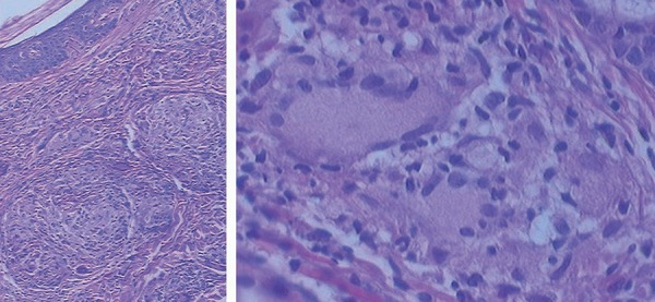
Sarcoidal granulomas on the dermis (left photo, hematoxylin and eosin, X100). Presence of multinucleated cells in the granuloma (right photo, hematoxylin and eosin, X400)

Sarcoidal granulomas on the dermis (left photo, hematoxylin and eosin, X100). Presence of multinucleated cells in the granuloma (right photo, hematoxylin and eosin, X400)