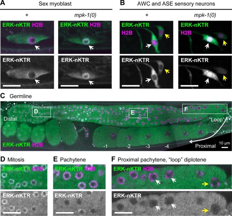
Figure 4. The ERK-nKTR reports MPK-1/ERK activity in multiple cell contexts.

(A) ERK-nKTR is excluded from nuclei of migrating Sex Myoblast (SM) cells in wild type, but not in mpk-1(0). SM-specific ERK-nKTR transgene arTi133, showing ERK-nKTR-mClover (ERK-nKTR) and mCherry-H2B (H2B) in wild type (+) and mpk-1(0). (B) ERK-nKTR is excluded from the nuclei of AWC and ASE sensory neurons in wild type, but not in mpk-1(0). AWC and ASE-specific ERK-nKTR transgene arTi137, showing ERK-nKTR-mClover (ERK-nKTR) and mCherry-H2B (H2B) in wild type (+) and mpk-1(0) AWC (white arrows) and ASE (yellow arrows) neurons. (C) ERK-nKTR is localized in a pattern correlated with MPK-1 activity in the hermaphrodite germline. Germline-expressed ERK-nKTR transgene arSi4, showing ERK-nKTR-mClover (ERK-nKTR) and mCherry-H2B (H2B). The most distal germline nuclei (oriented to left in this micrograph) are in mitosis. More proximally, nuclei enter meiosis and most of the length of germline remains in pachytene of Meiosis I. Proceeding proximally, the germline “loops”, and in this region nuclei enter diplotene, which is followed by diakinesis and oocyte maturation. (D-F) Images from insets indicated by boxes in (C). (D) ERK-nKTR accumulates in nuclei within the distal mitotic region, consistent with low MPK-1 activity detected in fixed samples (Lee et al., 2007). (E,F) ERK-nKTR levels are low in some nuclei in pachytene (E), but becomes highly excluded from nuclei in the proximal pachytene (white arrows) and loop region (yellow arrows) (F). In the most proximal region, ERK-nKTR is excluded from the nuclei undergoing diakinesis and oocyte maturation (C, labeled −1, −2, −3, −4). Nuclear exclusion of ERK-nKTR in proximal pachytene, diplotene, and diakinesis regions is consistent with regions where high levels of phosphorylated substrates are detected in fixed samples (Arur et al., 2011; Drake et al., 2014). Scale bars in all images are 10 μm.
