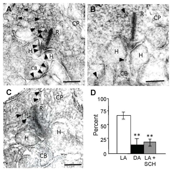
Figure 6. GABAAR expression on the dendritic tips of ON-cBCs is increased by bright light-evoked dopamine D1R activation.
(A–C) GABAARs were located at the plasma membrane of BC and HC dendritic tips following maintained bright illumination (A) but the number and size of clustered GABAAR-IR puncta were reduced following maintained darkness (B) and following maintained bright illumination when D1Rs were blocked with SCH (5 μM) (C). (D) Average percent of GABAAR-IR puncta in invaginations of ON-cBC dendritic tips at cone ribbon (triadic) synapses for each of the three experimental conditions. The average percent values shown were obtained by first determining the average percent value for each retina and then calculating the average value for all of the retinas in each experimental condition (1 retina/rabbit, 3 rabbits per experimental condition). GABAAR-IR was observed significantly more (p < 0.01; χ2 test for both LA vs. DA and LA vs. LA + SCH) in invaginated ON-cBC dendritic tips following maintained bright illumination (light-adapted, LA; total of 33 triadic synapses) than following maintained darkness (dark-adapted, DA; total of 27 triadic synapses) and following maintained bright illumination when D1Rs were blocked (LA + SCH; total of 23 triadic synapses). (A–C) Arrowheads: GABAAR-IR puncta; R: synaptic ribbons; CP: cone pedicles; CB: ON-cBC dendrites; H: HC dendrites; Scale bars: 100 nm. See also Figure S2.

