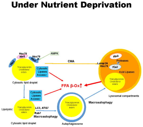
Figure 5. Schematic model of macroautophagy and chaperone-mediated autophagy (CMA) under conditions of nutrient deprivation.
Perilipins 2 and 3 provide a barrier to both cytosolic lipolysis and autophagy. Removal of perilipin (Plin) 2 and Plin 3 from the surfaces of lipid droplets is mediated by CMA and precedes lipolysis by both pathways. Plin 2 is phosphorylated by AMP kinase (AMPK) after associating with Hsc70. Plin 2 and Plin 3 are removed as complexes with Hsc70, followed by recognition by Lamp-2A on lysosomes, import into the lumen of the lysosome and degradation. Reduction of Plin 2 and Plin 3 on lipid droplets enables increased binding of cytosolic lipases (such as ATGL), Rab7 and protein effectors of macroautophagy (such as LC3-II) to lipid droplets. This leads to increased release of fatty acids by cytosolic lipases and the engulfment of the lipid droplet by the growing autophagosome. Autophagosomes fuse with lysosomes to form autolysosomes, wherein lysosomal acid lipases hydrolyze triacylglycerols and cholesterol esters, releasing fatty acids and cholesterol. Fatty acids are recycled back into the cytosol to mix with fatty acids generated by cytosolic lipolysis and support mitochondrial β-oxidation.

